Saturday, May 26, 2012

Diagnostic Yield of Fluoroscopy-Guided Biopsy for Infectious Spondylitis [SP...


Sent to you by subbu via Google Reader:


via Publication Preview by Kim, B. J., Lee, J. W., Kim, S. J., Lee, G. Y., Kang, H. S. on 5/24/12

BACKGROUND AND PURPOSE:

CT is currently the method of choice for guiding biopsy of lesions of the spine. However, in our hospital, fluoroscopy-guided percutaneous biopsy has been preferred for several years because of equipment availability and easy craniocaudal angulation. The aim of this study was to evaluate the efficacy of fluoroscopy-guided percutaneous biopsy in a clinical setting for diagnosing infectious spondylitis.

MATERIALS AND METHODS:

A retrospective study was performed to evaluate 170 fluoroscopy-guided percutaneous biopsies in 140 patients (male/female = 70:70; mean age, 65.1 years; range, 16–89 years) in a clinical setting who were suspected of having infectious spondylitis between July 2003 and March 2010. Diagnosis was based on pathologic confirmation by tissue or culture from biopsy. The percentage of adequate specimens for diagnosis, histopathologic diagnosis for infectious spondylitis, and positive cultures for causative organisms were evaluated by retrospective review of medical records.

RESULTS:

Adequate specimens for diagnosis were obtained in 165 of 170 cases (97.1%). The diagnosis of infectious spondylitis resulted in 134 of 170 cases confirmed through histopathology or clinical outcome (78.8%). In 51 of 134 cases (38.1%), the causative organism was confirmed by specimens from percutaneous bone biopsy. There were no biopsy-related major complications. The most common organism isolated was Mycobacterium tuberculosis (24 cases), followed by Staphylococcus aureus, Streptococcus agalactiae, and Streptococcus viridans.

CONCLUSIONS:

Fluoroscopy-guided percutaneous biopsy is as accurate and effective as CT-guided biopsy for diagnosing infectious spondylitis.


Things you can do from here:


CT Spotter: l5 spondylolysis

image

image

image

Venous thrombosis presenting as subarachnoid hemorrhage

49 yrs, M

Seizures on the morning of admission

Vitals stable

No focal deficit

A CT done at admission showed SAH with flocculent surface hematoma in the left anterior frontal region…significantly there was no blood in the basal cisterns

image

NCCT at admission

image

image

Enlarged images of the NCCT

A cerebral DSA was done which showed thrombosis of anterior part of the superior sagittal sinus, partial thrombosis of the inferior sagittal sinus with hypoplastic left transverse and sigmoid sinuses

image

image

image

Right ICA angiogram

image

image

Left ICA angiogram

image

Left and right vertebral angiogram

image

Right middle meningeal had anomalous origin from right petrous ICA

image

image

Left ECA angiogram showed blush from the middle meningeal artery in the region of the thrombosed superior sagittal sinus

Patient was put on heparin after this

image

NCCT next morning showed significant resolution of the SAH.

image

image

Enlarged images of the NCCT

The patient had an uneventful recovery and was discharged in a stable condition, on Warfarin.

Tuesday, May 22, 2012

Giant Sacral Schwannoma

This is the case of a female patient with giant sacral neurogenic tumor. The patient was remarkably preserved for the tumor size and mass was found on ultrasound for gynecological evaluation.

Friday, May 11, 2012

Ankylosing spondylitis with cervical spine fracture

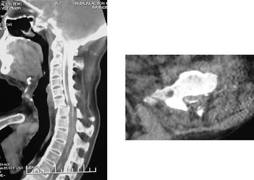
A 51 years old male patient, known case of ankylosing spondylitis since many years, had severly curved totally fused spine. He suffered a minor fall followed by weakness of limbs. Due to the curvature, he did not fit in the MRI machine. A CT was done which showed frature of the lower cervical spine with displacement. CT myelogram was then tries but could not be done from the lumbar site as the needle could not be passed through the heavily calcified tissue. Then the needle was inserted through the suboccipital route and contrast injected to perform the CT-myelo.

The myelogram showed partial obstruction to the contrast flow however the compression was not extremely severe. The patient was operated and the spine fixed.

Twitter Delicious Facebook Digg Stumbleupon Favorites More

 
Design by Free WordPress Themes | Bloggerized by Lasantha - Premium Blogger Themes | Blogger Templates