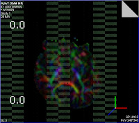
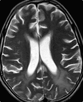
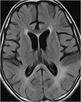
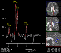
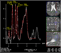
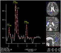
Conventional MRI of a 30 years old male patients with intractable right sided partial seizures showed the presence of left superior frontal cortical dysplasia
Tractography showed the unorganised white matter bundles in the left frontal lobe
However it unravelled more extensive changes of white matter disorganisation invvelving almost whole of the left hemisphere.
Tractography also revealed an area of absence of fibres in the left lower thalamus, subthalamic paraventricular region probable related to an underlying lesion there-? heterotopiaFig 1. color coded tracts overlayed upon MPRAGE image at centrum semiovale level (side reversed): shows 'more' number of fibres in the left side. The tracts are unorganised as evident by the haphazard intemingling of the different colors. The bulge towards the midline anteriorly is the area seen on conventional MRI. Note that the above-said area has maximum disorganisation(lot of mixture of purple, red, blue,green and yellow)
The following images (Side reversed) shows the focal 'void' in the tract bundles in the left inferior thalamus paraventricular region
The color coded DTI map (side unreversed)shows the same finding. Note that the area of 'void' in the tractography images seen above shows a collection bunch of diferent colors ( mainly purple) indicating a loss of coherent anisotropy


 1:47:00 PM
 Dr Subhash Kumar, MBBS, MD, DM


