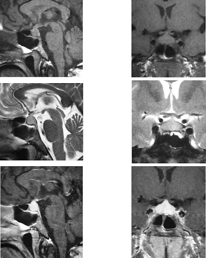
A middle aged male had undergone an MRI brain for repeated headaches.

There was diffuse enlargement of the pituitary with thickening of the stalk, the T1 - bright appearing posterior pituitary was however seen separately. The enlarged pituitary was isointense on T1 and bright on T2 images with intense enhancement on post-Gd images. There was no other intracranial pathology detectable.
CSF examination was unremarkable, so were other general tests.
The patient underwent endoscopic surgery. There were adhesions all around and the mass could be removed with great diffuculty. Intra-op there was CSF leak which was sealed.
After surgery patient developed diabetes insipidus and after 7days he died.
Histopathologic exam showed granulomas and the diagnosis was granulomatous hypophysitis.


 11:25:00 AM
 Dr Subhash Kumar, MBBS, MD, DM
 Posted in:
