Wednesday, November 18, 2009

A strange case of Tuberculosis

A 27 years old man presented with seizures and left sided hemiparesis

DSC01418

The CT shows hypodensity in right temporo-parieto-occipital region

An MRI done a month later shows extensive nodular enhancing lesions with lot of edema. Significantly the enhancing area were T1 bright and T2 hypointense.

DSC01415 DSC01417 DSC01414

After starting antitubercular treatment the edema and the enhancement subsided.

But the patient continues to have seizures and weakness persists.

The 4 month follow up CT:

DSC01406

Friday, November 13, 2009

A young man with vision loss: Adrenoleukodystrophy

DSC02213

DSC02218  DSC02214

DSC02222DSC02219

A 19 years old male, otherwise healthy presented with gradually increasing vision loss and weakness of both both lower limbs and normal routine laboratory investigations. His MRI revealed confluent bilateral (L>R) parieto-occipital signal change, tissue loss, inflammatory enhancing edge s/o ALD

Wednesday, October 7, 2009

An elderly gentleman with inability to hold urine

87 years old male with Gradually worsening bladder symptoms—unable to hold urine, Emotional lability,Occasional falls-unsteadiness while walking since last 10 months


Diagnosis: Normal Pressure hydrocephalus
Findings: Triventricular dilatation out of proportion to atrophy with periaqueductal hyperintensity, large jet of flow void in the aqueduct and fourth ventricle, periventricular hyperintensity, upward ballooning of corpus callosum, posterior gyri opposed aginst the calvarium, CSf pocket in medial sulcus
Pre CSF tap CSF flow study result through aqueduct shows peak veocity of 16.92cm/sec and stroke volume of 111 mirolt/sec while post CSF tap (70 ml) CSF flow study sows peak velocity of 7.79 cm/sec and stroke volume of 8 microlt/sec.

Wednesday, July 8, 2009

Tractography finds unseen lesions in a case of cortical dysplasia!

Conventional MRI of a 30 years old male patients with intractable right sided partial seizures showed the presence of left superior frontal cortical dysplasia
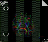
Tractography showed the unorganised white matter bundles in the left frontal lobe
However it unravelled more extensive changes of white matter disorganisation invvelving almost whole of the left hemisphere.
Tractography also revealed an area of absence of fibres in the left lower thalamus, subthalamic paraventricular region probable related to an underlying lesion there-? heterotopia

Fig 1. color coded tracts overlayed upon MPRAGE image at centrum semiovale level (side reversed): shows 'more' number of fibres in the left side. The tracts are unorganised as evident by the haphazard intemingling of the different colors. The bulge towards the midline anteriorly is the area seen on conventional MRI. Note that the above-said area has maximum disorganisation(lot of mixture of purple, red, blue,green and yellow)
The following images (Side reversed) shows the focal 'void' in the tract bundles in the left inferior thalamus paraventricular region
The color coded DTI map (side unreversed)shows the same finding. Note that the area of 'void' in the tractography images seen above shows a collection bunch of diferent colors ( mainly purple) indicating a loss of coherent anisotropy


Tuesday, July 7, 2009

Chronic Multiple Sclerosis

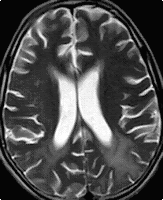
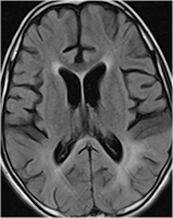
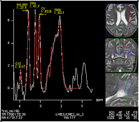
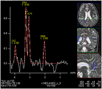
A case of chronic RRMS shows diffuse atrophy with periventricular white matter hyperintensity
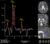
MR spectroscopy from left parieto-occipital white matter, which has the maximum hyperintensity, shows, as expected raised choline, decreased NAA and presence of lactate, i.e changes of demyelination and tissue loss
MRS from right parieto-occipital white matter also shows decreased NAA/Cho ratio and raised lactate


However, noteworthy is the presence of similar changes on spectroscopy in the normal appearing areas too


Saturday, July 4, 2009

Tractography in right frontal glioma


Preoperative tractography done in a large frontal lobe anaplastic astrocytoma.
Look at the 'absence' of the white matter tracts.
Some tracts have been displaced and stretched but a significant part of the white matter has disappeared altogether.

Twitter Delicious Facebook Digg Stumbleupon Favorites More

 
Design by Free WordPress Themes | Bloggerized by Lasantha - Premium Blogger Themes | Blogger Templates