Saturday, May 26, 2012

Diagnostic Yield of Fluoroscopy-Guided Biopsy for Infectious Spondylitis [SP...


Sent to you by subbu via Google Reader:


via Publication Preview by Kim, B. J., Lee, J. W., Kim, S. J., Lee, G. Y., Kang, H. S. on 5/24/12

BACKGROUND AND PURPOSE:

CT is currently the method of choice for guiding biopsy of lesions of the spine. However, in our hospital, fluoroscopy-guided percutaneous biopsy has been preferred for several years because of equipment availability and easy craniocaudal angulation. The aim of this study was to evaluate the efficacy of fluoroscopy-guided percutaneous biopsy in a clinical setting for diagnosing infectious spondylitis.

MATERIALS AND METHODS:

A retrospective study was performed to evaluate 170 fluoroscopy-guided percutaneous biopsies in 140 patients (male/female = 70:70; mean age, 65.1 years; range, 16–89 years) in a clinical setting who were suspected of having infectious spondylitis between July 2003 and March 2010. Diagnosis was based on pathologic confirmation by tissue or culture from biopsy. The percentage of adequate specimens for diagnosis, histopathologic diagnosis for infectious spondylitis, and positive cultures for causative organisms were evaluated by retrospective review of medical records.

RESULTS:

Adequate specimens for diagnosis were obtained in 165 of 170 cases (97.1%). The diagnosis of infectious spondylitis resulted in 134 of 170 cases confirmed through histopathology or clinical outcome (78.8%). In 51 of 134 cases (38.1%), the causative organism was confirmed by specimens from percutaneous bone biopsy. There were no biopsy-related major complications. The most common organism isolated was Mycobacterium tuberculosis (24 cases), followed by Staphylococcus aureus, Streptococcus agalactiae, and Streptococcus viridans.

CONCLUSIONS:

Fluoroscopy-guided percutaneous biopsy is as accurate and effective as CT-guided biopsy for diagnosing infectious spondylitis.


Things you can do from here:


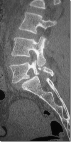
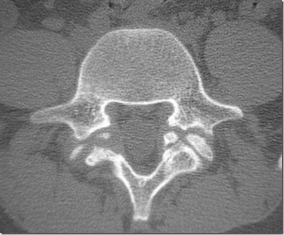
CT Spotter: l5 spondylolysis

image

image

image

Venous thrombosis presenting as subarachnoid hemorrhage

49 yrs, M

Seizures on the morning of admission

Vitals stable

No focal deficit

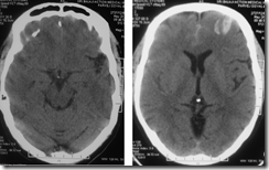
A CT done at admission showed SAH with flocculent surface hematoma in the left anterior frontal region…significantly there was no blood in the basal cisterns

image

NCCT at admission

image

image

Enlarged images of the NCCT

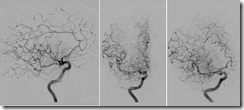
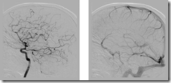
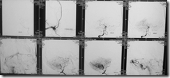
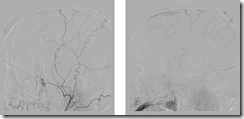
A cerebral DSA was done which showed thrombosis of anterior part of the superior sagittal sinus, partial thrombosis of the inferior sagittal sinus with hypoplastic left transverse and sigmoid sinuses

image

image

image

Right ICA angiogram

image

image

Left ICA angiogram

image

Left and right vertebral angiogram

image

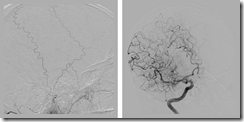
Right middle meningeal had anomalous origin from right petrous ICA

image

image

Left ECA angiogram showed blush from the middle meningeal artery in the region of the thrombosed superior sagittal sinus

Patient was put on heparin after this

image

NCCT next morning showed significant resolution of the SAH.

image

image

Enlarged images of the NCCT

The patient had an uneventful recovery and was discharged in a stable condition, on Warfarin.

Tuesday, May 22, 2012

Giant Sacral Schwannoma

This is the case of a female patient with giant sacral neurogenic tumor. The patient was remarkably preserved for the tumor size and mass was found on ultrasound for gynecological evaluation.

Friday, May 11, 2012

Ankylosing spondylitis with cervical spine fracture

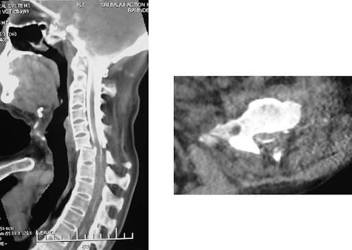
A 51 years old male patient, known case of ankylosing spondylitis since many years, had severly curved totally fused spine. He suffered a minor fall followed by weakness of limbs. Due to the curvature, he did not fit in the MRI machine. A CT was done which showed frature of the lower cervical spine with displacement. CT myelogram was then tries but could not be done from the lumbar site as the needle could not be passed through the heavily calcified tissue. Then the needle was inserted through the suboccipital route and contrast injected to perform the CT-myelo.

The myelogram showed partial obstruction to the contrast flow however the compression was not extremely severe. The patient was operated and the spine fixed.

Granulomatous hypophysitis

A middle aged male had undergone an MRI brain for repeated headaches.

There was diffuse enlargement of the pituitary with thickening of the stalk, the T1 - bright appearing posterior pituitary was however seen separately. The enlarged pituitary was isointense on T1 and bright on T2 images with intense enhancement on post-Gd images. There was no other intracranial pathology detectable.

CSF examination was unremarkable, so were other general tests.

The patient underwent endoscopic surgery. There were adhesions all around and the mass could be removed with great diffuculty. Intra-op there was CSF leak which was sealed.

After surgery patient developed diabetes insipidus and after 7days he died.

Histopathologic exam showed granulomas and the diagnosis was granulomatous hypophysitis.

Thursday, May 10, 2012

CLOTBUST sonothrombolysis therapy for acute left MCA territory stroke

CLOTBUST sonothrombolysis in acute stroke is a new addition in the armamentarium of physicians traeting these particularly challenging patients. It uses the simple premise of continuous transfer of ultrasonic wanes to the site of the clot in the blood vessel, which in a way soften ups the clot, such that intravenously given thrombolytic agents can work more effectively, resulting in better recanalisation rates, hence translating into better patient outcome.
We recently performed it on a patient with 3.5 hours of acute stroke onset, patient presenting with right sided weakness and Wernicke's aphasia, MRI showing acute left MCA territory diffusion restriction. As the DWI volume was not much and ASPECTS score was good, thrombolysis was performed along with continuous transcranial Doppler using a 2MHz probe, for two hours. After about 45 minutes, the patient responded, sat up himself and the weakness improved. Over the next 48 hours he continued to improve and had 4+/5 power with some aphasia; the aphasis improved over next two weeks and the patient is independent for ADL.
The TCD frame with the doppler probe in place

MRI showing diffusion restriction in left MCA territory


I started the doppler
The TCD wave at the end of the procedure ( 2 hours)
The patient sat up on his own


Simple coiling of a wide necked aneurysm


Wide necked aneurysms often are deemed to require assistance with balloon or stent during endovascular coiling, however, most of them do not require support as such, and well placed coils do the job.
Here is an example, wherein a wide neck Acom aneurysm incorporating one of the A2 segments, was coiled well without use of any balloon/stent.


Friday, April 27, 2012

Cases in this blog....click on the link to open the full case page

  1. PET picks up Rasmussen's encephalitis before CT/MRI: http://neuroradiologyindia.blogspot.in/2010/06/pet-picks-up-rasmussens-encephalitis.html
  2. A strange case of tuberculosis: http://neuroradiologyindia.blogspot.in/2009/11/strange-case-of-tuberculosis.html
  3. A young man with vision loss: Adrenoleukodystrophy: http://neuroradiologyindia.blogspot.in/2009/11/young-man-with-vision-loss.html
  4. An elderly gentleman with inability to hold urine: normal pressure hydrocephalus: http://neuroradiologyindia.blogspot.in/2009/10/urine.html
  5. Tractography finds unseen lesions in a casae of cortical dysplasia: http://neuroradiologyindia.blogspot.in/2009/07/tractography-finds-unseen-lesions-in.html
  6. Chronic multiple sclerosis: http://neuroradiologyindia.blogspot.in/2009/07/chronic-multiple-sclerosis.html
  7. Tractography in right frontal glioma: http://neuroradiologyindia.blogspot.in/2009/07/tractography-in-right-frontal-glioma.html

Thursday, April 26, 2012

Gunshot injury to cervical spine and vertebral artery

A young man had accidentally shot himself, the bullet injuring his face, breaking the mandible and lodging on the right side of the cervical spine.

The CT images show the foreign body impinging the right foramen transversarium of the third cervical vertebra.

For evaluating the vertebral artery,a DSA was planned.


The DSA showed slight narrowing of the right vertebral artery, but with normal antegrade flow; lucky shave for the patient as it was the dominant artery and the left vertebral ended in the PICA.

The patient was put on conservative management.

Monday, April 23, 2012

Tetralogy of Fallot With Midbrain Abscess

High Grade Glioma With Metastases

Acute Stroke IV Thrombolysis

Multiple Myeloma

Pinealoblastoma

CADASIL vs HYPERTENSIVE MICROANGIOPATHY

Multifocal anaplastic oligoastrocytoma

Phenylketonuria vs perinatal insult

A confusing case of a boy from middle east presenting with developmental delay. He had been diagnosed as phenylketonuria on lab investigations, plus he had blue eyes and light hairs. However clinically he did not seem to fit into it. Hence investigations were repeated and this time they were normal.

Perinatal Insult

Twitter Delicious Facebook Digg Stumbleupon Favorites More

 
Design by Free WordPress Themes | Bloggerized by Lasantha - Premium Blogger Themes | Blogger Templates